Ok, so. Keeping in mind that my brain is absolutely mush from being at overcapacity, and the fact that I am enjoying what I feel is a rather deserved glass of wine at the moment, I am going to do my best to explain what we think is going on. Sorry in advance for the long post, though I promise this is the condensed version. You want a summary? Skip to the bottom.
[First things first- you remember Foster’s diary that I keep? That has been invaluable during this whole process, and if your type A-ness needs an outlet and you have an accident prone horse, I highly recommend it.]
The Bone Scan
Let’s start with the bone scan. The bone scan really frustrated me at first, in that the results seemed inconclusive to little-old-not-a-veterinarian-me. Basically, the areas that “lit up” (actual term: increased radiopharmaceutical uptake) were his hocks, slightly FL navicular area (which had me saying WTF- that’s the good leg), and slightly outside cannon bone HL. That troubled FR, and really lame HR? Nada.
But from the bone scan we were able to rule out anything really active in the FR, the stifles, and the SI area (which we thought would be the problem). The hocks, after getting 2 radiologists’ opinions, are likely bone edema/remodeling and though a source of discomfort, not likely to cause the acute lameness we are seeing.

The Lameness Exam
Fast forward 3 days to tonight’s vet appointment. We had chatted yesterday about the results, and were thinking probably suspensory issue in the RH but also the possibility (though small) of neurological disease such as EPM or Lyme. So the plan was to ultrasound the leg and/or do nerve blocks to pinpoint the lameness.
We trotted him up the concrete aisle and threw him on the lunge to get a baseline lameness, and saw that the RF seems to have resolved itself, and we were still looking at a definite RH, slight LH lameness. After much discussion, we decided to ultrasound the leg first and then nerve block after (not the typical way of doing things, but I was getting anxious). But first we would block the foot just in case, since that wouldn’t interfere with the ultrasound and we could rule it out if it didn’t improve.
And what do you know, it looked better. A lot better. Now knowing that the lameness was in the foot, we could consider 3 causes:
- The palmar angle is too low and is making him extremely heel sore
- Deep digital flexor tendon injury
- Injury to any of the ligaments in and around the navicular bone
So we blocked the left hind foot, since that was also still showing lameness.
And that looked better, too.

More walking
In Summary
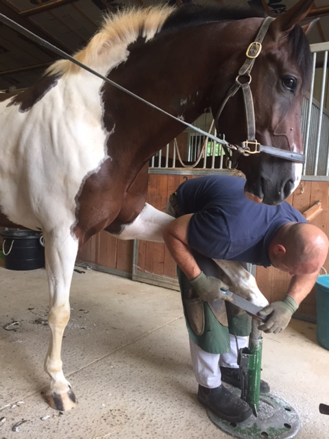
So, while we don’t have a definitive answer, we have a location(s) on the body to concentrate on. The vet thinks that the palmar angles in the hind feet are likely the key to all this, and so we will be doing X-Rays and having a discussion with my farrier a week and a half from now, basically as soon as we can squeeze him in. Ideally the X-Rays will confirm terrible palmar angles and we can begin with corrective shoeing to sort it out.
It may take as long as two shoeing cycles to allow Foster’s feet time to heel and react to the changes, in which time I can work him but must keep all concussion to his feet at a minimum- in other words, walk only. I can do whatever I want at the walk, which is great news to me, but trotting should be at a minimum and cantering and jumping are definitely out. If, after those 2 shoeing cycles, we bring him back in to work and he is no better or still lame, then we will have to look at getting an MRI done to determine soft tissue injury. I would also assume that if the X-Rays show awesome palmar angles, an MRI will be more quickly in our future.
I can’t tell you how relieved I am just to even know where the problem is, and even more so to be hopeful that it can be resolved with corrective shoeing. I’m actually hoping to see some pretty terrible x-rays at our next farrier appointment, and I’ll certainly be posting an update when I have it.
Until then- bring on the walk suggestions! What fun walk exercises can we do?


 Right now we’re playing the waiting game until the radiography specialist interprets the images, which I’m told could be later the afternoon or even tomorrow. And then, once we get those results, I’ll be speaking with my vet to determine a plan of action.